OPS>RESEARCH>POLARIZED LIGHT FIELD
Polarized Light Field Microscopy (Oldenbourg Lab)
Current microscopy methods supported by OpenPolScope technology are orthoscopic, i.e., they image the anisotropy of a specimen as it appears along a single viewing direction, usually the microscope optical axis. For example, if a birefringent crystal is oriented with its optic axis parallel to the microscope axis, the crystal appears isotropic, or non-birefringent. Thus, orthoscopic imaging methods provide an incomplete description of the specimen's anisotropy, unless several views can be examined by rotating the specimen in 3D using a universal stage (Images of a Fedorov universal stage). The universal stage, however, severely reduces achievable image resolution, as it requires long working distance objective lenses.
The traditional method of conoscopic imaging with the polarizing microscope achieves the analysis along different viewing directions by examining the back focal plane of the microscope objective lens. However, conoscopic imaging requires restricting the illumination to a small specimen region occupied by a single crystal, for example. Only then can the distribution of light intensity and its polarization in the back focal plane reveal the crystal's anistropy, one crystal at a time.
The new approach of polarized light field microscopy promises to exploit the angular diversity of microscopic imaging to create a multitude of views of the specimen, while maintaining most of the spatial resolution. The multitude of views are generated by the placement of a light field sensor in the image plane of the microscope objective lens, without changing the optical path of the microscope.
A light field sensor consists of a microlens array followed by a CCD or similar detector array. The detector array is placed in the focal plane of the lenslets, so that parallel rays impinging on a lenslet from a certain direction are focused into a single detector pixel. The microlens array has typically 200 x 200 lenslets and behind each lenslet are 10 x 10 detector pixels. Hence, the output of the detector array consists of 200 x 200 spatial samples, each consisting of 10 x 10 directional samples, for light that has traveled through the specimen. The combination of spatial and directional information is called a light field.
We have built a prototype Light Field OpenPolScope and published first results by examining the birefringence of a thin layer of calcite crystals: Polarized light field microscopy: an analytical method using a microlens array to simultaneously capture both conoscopic and orthoscopic views of birefringent objects.
 The image to the right was recorded with the Birefringence OpenPolScope and displays the retardance (brightness) and slow axis orientation (hue) of a 300 nm thick layer of calcite crystals. Each crystalline domain has nearly uniform hue and brightness, due to the uniform birefringence of each domain. Variation in brightness between domains are caused by differences in the inclination angle of the optic axis of each domain. The inclination angle of the optic axis of a domain becomes apparent in the light field image below.
Images acquired using a 63/1.4 NA objective and 1.4 NA condenser lens (both oil immersion lenses).
 The same calcite layer imaged with the Birefringence OpenPolScope equipped with a light field sensor. In the enlarged region, a crystal reveals the orientation of its optic axis by the retardance pattern measured behind each microlens. For example, light that has traversed the dark crystal domain generates a circular retardance pattern with an azimuthal slow axis orientation behind each microlens. The central dark spot represents the direction of the optic axis of the crystal (see high-resolution version of image).
Prototype of polarized light field microscope
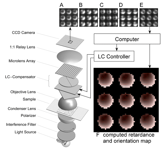
On the right, we show a schematic of the polarized light field microscope including the optical set-up with image acquisition and processing components. The optical parts include the light source (100W halogen lamp), interference band pass filter (wavelength 546 nm/10 nm band pass), left circular polarizer, condenser lens (oil, NA 1.4), sample (calcite film), objective lens (PlanApo 63x/1.4 NA oil), universal compensator made of 2 liquid crystal variable retarder plates and a linear polarizer, microlens array (placed in the objective's image plane), and a 1:1 relay lens, which images the back focal plane of the microlenses onto the CCD sensor. (A–E) Complete analysis of the polarized light field requires 5 images; each image shown is a 52×52 sub-region of the five 2048×2048 pixel images actually acquired, each image recorded at one of the five settings of the LC-compensator. Based on the raw intensity images, the computer calculates a retardance and an orientation map, shown here as a composite image with red lines indicating the slow axis orientation. Each of the 9 disk-shaped images represent a multitude of views of a small sample area. Dark centers of the circular patterns represent the viewing direction along the optic axis of the calcite crystal.
Current Research
In a recent publication, we examine the point spread function of the polarized light field microscope and establish a computational approach to solve the forward problem in polarized light field imaging. We recorded experimental polarized light field images of small calcite crystals and of larger birefringent objects and compare our experimental results to numerical simulations based on polarized light ray tracing. We find good agreement between all our experiments and simulations, which leads us to propose polarized light ray tracing as one solution to the forward problem of imaging with the polarized light field microscope. Solutions to the ill-posed, non-linear inverse problem might be found in analytical methods and/or deep learning approaches that are based on training data generated by the forward solution presented in this publication.
The simulation code is available as a Mathematica Notebook and is an open-source companion piece to the forthcoming article. For more information click here.
Marc Levoy, Stanford University
Light field photography is an image-based technique for recording scene appearance. Unlike conventional photography, light fields permit manipulation of viewpoint and focus after the imagery has been recorded. Devices for capturing light fields range in scale from room-size arrays of cameras to a handheld camera in which a microlens array has been inserted between the main lens and sensor plane. Read more at: Stanford Light Field Microscope Project
Light Field Photography, Microscopy and Illumination, Levoy
Light field micrographs captured at MBL

